Course Detail
Environmental Microbiology
List of Contents
Water Microbiology
Drinking water or potable water, ideally, should not only be safe but also pleasant to drink. It should be clear, colorless, and devoid of disagreeable taste or smell. It should be free from pathogenic microorganisms and chemical substances.
Many people struggle to obtain access to safe water. A clean and treated water supply to each house may be the norm in Europe and North America, but in developing countries, access to both clean water and sanitation are not the rule, and waterborne infections are common. Two and a half billion people have no access to improved sanitation, and more than 1.5 million children die each year from diarrheal diseases. According to the WHO, the mortality of water associated diseases exceeds 5 million people per year. From these, more that 50% are microbial intestinal infections, with cholera standing out in the first place. In general terms, the greatest microbial risks are associated with ingestion of water that is contaminated with human or animal feces. Wastewater discharges in fresh waters and costal seawaters are the major source of fecal microorganisms, including pathogens.
Bacterial flora in water can be classified into three groups as follows:
- Natural water bacteria: These are the bacteria that are commonly found in water free from gross pollution.
- Soil bacteria: These are the bacteria that are not normal inhabitants of water but are found after being washed into the water during heavy rains.
- Sewage bacteria: These are the bacteria that are not normal inhabitants of water but are found in water after being contaminated with sewage. These bacteria include those which are the normal inhabitants of the intestine of humans and animals. These also include the bacteria that live mainly on decomposed organic matter of either plant or animal origin.
The following factors determine the number of bacteria in water:
- Salinity: The number of bacteria present in water depends on salinity of water; in most cases, the more the salinity, the less the number of bacteria.
- Acidity: Acidity of water has a deleterious effect on most of the bacteria.
- Temperature: Low temperature usually favors survival of the bacteria. When temperatures are below freezing, the growth of bacterria may be inhibited, but this growth resumes when temperatures rise above freezing.
- Light: Sunlight with the wavelength of 300–400 nm is highly bactericidal, provided water is clear and static. The bactericidal effect is reduced due to the presence of organic matter and due to movement in water.
- Storage: Storage of water decreases bacterial count in stored water due to sedimentation and revitalization.
- Organic matter: When organic matter is plenty, the microorganisms tend to multiply and are present in large numbers whereas when it is less, the organisms are few. organic matter also tends to inhibit the efficacy of disinfectants and other chemicals so the bacteria numbers increase.
- Type of water: Surface water is more likely to be contaminated than the deep water. The latter is usually pure.
Indicator Organisms
The presence of indicator organisms indicates that fecal matter has entered the water supply, or the fecal bacteria have not been killed or removed by purification processes, and/or the water supply, therefore, is liable to be contaminated with dangerous intestinal pathogens.
Some of the organisms that are used as indicator organisms are Coliforms, Fecal streptococci and Sulfite-reducing clostridia.
Collection of water samples for analysis
Sampling from a tap or pump outlet: When collecting from taps, allow the water to run to waste for 2-3 minutes before running it into the bottle. Then the stopper of the bottle is opened, it is filled with water, and the stopper is replaced.
Sampling from a reservoir, such as stream, river, lakes, and tanks: When sampling from streams or lakes, the bottle is opened at a depth of about 30 cm with its mouth facing the current and it is ensured that the water entering the bottle has not been in contact with hands.
Sampling from a well: The sampling bottle is tied with a stone and a clean cord of suitable length and is lowered to the required depth in the well. The bottle is completely immersed in the water. When the bottle is completely filled, it is pulled out and then it is stoppered. It is ensured that the bottle does not touch the side of the well at any time.
Methods of water analysis
Following tests are generally done for the routine bacteriological analysis of water:
- Presumptive coliform count
- Differential coliform count
- Membrane filtration method
- Detection of fecal streptococci and C. perfringens
- Detection of specific pathogens
The main bacterial diseases transmitted through drinking water.
- Cholera: Vibrio cholerae, serovarieties O1 and O139.
- Gastroenteritis caused by vibrios: Vibrio parahaemolyticus.
- Typhoid fever and other serious salmonellosis: Salmonella enterica subsp. enterica serovar Paratyphi, serovar Typhi and serovar Typhimurium.
- Bacillary dysentery or shigellosis: Shigella dysenteriae, Shigella flexneri, Shigella boydii and Shigella sonnei.
- Acute diarrheas and gastroenteritis: Escherichia coli, particularly serotypes such as O148, O157 and O124.
Cholera
The incubation period is short and varies from 2 to 3 days after ingestion of the bacteria. The condition shows an abrupt onset of watery diarrhea and vomiting. Profuse watery diarrhea is the most important manifestation of cholera. The volume of diarrheic stool excreted in cholera is much more than that of diarrhea caused by any other infectious pathogen. In a severe condition, patients may excrete as high as 250 mL of stool per kg body weight in a day.
Severe abdominal cramp, possibly caused by distension of the small intestine due to excretion of larger volumes of intestinal fluid, is also seen in these patients. Vomiting is another important manifestation of cholera and occurs in early stage of the disease. This is caused by decreased gastric and intestinal motility.
Dehydration in cholera characteristically develops very fast, within hours after the onset of symptoms. This rapid development of dehydration is not seen with diarrheal diseases caused by any other enteropathogen. In untreated patients, case fatality rate has been estimated to vary from 25% to 50%. In patients treated well with replacement of lost fluid and electrolytes, the case fatality is usually less than 1%.
Epidemiology
Cholera continues to be a major health problem in many parts of the world including Indian subcontinent and sub- Sahara in Africa. The condition is rare in the developed and industrious nations for the last many decades.
Habitat: V. cholerae is a salt water bacterium. Marine ecosystem in association with plankton is the primary habitat of the bacteria. The bacteria can multiply freely in the water
Diagnosis
Fresh stool specimen collected before administration of antibiotics is the specimen of choice. This can be followed with microscopy or culture.
Serotyping: using specific V. cholerae O1 antisera. In this test, the colonies are picked up with a straight wire and mixed with a drop of antisera on the slide. Agglutination of the bacteria shows that the test is positive for V. cholerae O1.
Treatment
Treatment of cholera includes (a) replacement of fluid and electrolytes and (b) antibiotic therapy.
Prevention
The preventive measures against cholera include general preventive measures and cholera vaccination.
Vibrios that require a higher concentration of sodium chloride are known as halophilic vibrios. They are natural inhabitants of sea water and marine life. V. parahaemolyticus,Vibrio alginolyticus, and V. vulnificus are three important halophilic vibrios species known to cause infection in humans.
V. parahaemolyticus is now recognized as an important cause of seafood-associated gastroenteritis throughout the world.
V. alginolyticus is widely distributed in sea water and seafood. V. alginolyticus is associated with infections of superficial wounds exposed to contaminated sea water.
V. vulnificus is a more virulent bacterium than V. parahaemolyticus. Presence of capsule and production of hydrolytic enzymes, such as cytolysins, proteases, and collagenases are responsible for virulence of the bacteria.
Salmonellosis
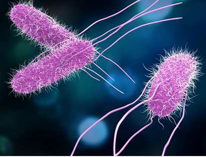
Salmonella is a genus of rod-shaped (bacillus) Gram-negative bacteria of the family Enterobacteriaceae. The two species of Salmonella are Salmonella enterica and Salmonella bongori. Salmonella was named after Daniel Elmer Salmon, an American veterinary surgeon (1850-1914).
Salmonella species are non-spore-forming, predominantly motile enterobacteria with cell diameters between about 0.7 and 1.5 µm, lengths from 2 to 5 µm, and peritrichous flagella.
Culture
Depending on the environmental conditions, Salmonella spp can be chemotrophs, (obtaining their energy from oxidation and reduction reactions using organic sources), facultative anaerobes, (capable of generating ATP with oxygen ("aerobically") when it is available), or using other electron acceptors or fermentation ("anaerobically") when oxygen is not available.
They grow at an optimum temperature of 37oC in a pH of 6-8 on a variety of nonselective (Mueller-Hinton agar) and selective (Wilson and Blair's bismuth sulfite medium) media.
Biochemical Reactions
Salmonellae show following reactions:
- They ferment glucose, mannitol, and maltose, forming acid and gas. S. Typhi is an exception, which does not ferment the sugars.
- They do not ferment lactose, sucrose, or salicin.
- They do not produce indole.
- Most salmonellae except S. Paratyphi A, S. Choleraesuis, and some other species produce H2S.
- They do not hydrolyze urea. They are MR positive and VP negative and citrate positive.
The bacilli are killed at a temperature of 55oC in 1 hour or at 60oC in 15 minutes. They are also killed by 0.2% mercuric chloride or 5% phenol in 5 minutes. Boiling, chlorination of water, and pasteurization of milk kill the bacteria.
Pathogenesis and Immunity
Salmonella possess three major antigens:
1. H or flagellar antigen: This antigen is present on the flagella
and is heat and alcohol labile.
2. O or somatic antigen: O antigens occur on the surface of
the outer membranes and are determined by specific sugar
sequences on the cell surface.
3. Surface antigens (Vi antigen, M and N antigen, and F antigens)
Salmonella spp are intracellular pathogens. Most infections are due to ingestion of food contaminated by animal feces, or by human feces, such as by a food-service worker at a commercial eatery. Individuals with depressed CMI appear to be more susceptible to S. Typhi infections. In acute infection, O antibody appears first, rising progressively, later falling, and often disappearing within a few months; H antibody appears slightly later but persists longer. Rising or high O antibody titers generally indicate acute infection, whereas elevated levels of H antibody help to identify the type of enteric fever.
The severity of disease in individuals infected with salmonellae is dependent on the virulence factors of the infecting strain as well as on the human host. Salmonella serotypes can be divided into two main groups-typhoidal and nontyphoidal. On reaching the intestine, the salmonellae attach themselves by fimbriae or pili to cells lining the ileal mucosa.
Typhoidal serotypes can only be transferred from human-to-human, and can cause food-borne infection, typhoid fever, and paratyphoid fever. Typhoid fever is caused by Salmonella invading the bloodstream (the typhoidal form), or in addition spreading throughout the body, invading organs, and secreting endotoxins (the septic form). This can lead to life-threatening hypovolemic shock and septic shock, and requires intensive care including antibiotics. Nontyphoidal serotypes can be transferred from animal-to-human and from human-to-human. They usually invade only the gastrointestinal tract and cause salmonellosis, the symptoms of which can be resolved without antibiotics. However, in sub-Saharan Africa, nontyphoidal Salmonella can be invasive and cause paratyphoid fever, which requires immediate treatment with antibiotics.
Because Salmonella are intracellular, cell mediated immunity (CMI) rather than humoral antibodies play more important role in protection against the disease.
A summary of diseases caused by Salmonella species is shown below:
| Salmonella species | Disease/syndrome |
|---|---|
| Salmonella Typhi | Typhoid fever |
| Salmonella Paratyphi | Paratyphoid fever |
| Salmonella Cholerasuis | Salmonella bacteremia |
| Salmonella Typhimurium | Salmonella gastroenteritis |
| Salmonella Enteritidis | Salmonella gastroenteritis |
| Salmonella Hadar | Salmonella gastroenteritis |
| Salmonella Heidelberg | Salmonella gastroenteritis |
| Salmonella Agona | Salmonella gastroenteritis |
| Salmonella Virchow | Salmonella gastroenteritis |
| Salmonella Seftenberg | Salmonella gastroenteritis |
| Salmonella Indiana | Salmonella gastroenteritis |
| Salmonella Newport | Salmonella gastroenteritis |
| Salmonella Anatum | Salmonella gastroenteritis |
S. Typhi is an invasive bacterium. It colonizes the human intestine and, under specific conditions, directly invades the intestinal mucosa or multiplies for several days within the mononuclear phagocytic cells in the liver, spleen, lymph nodes, and Peyer patches of the ileum before invasion.
Epidemiology
Enteric fever is endemic in many developing countries especially where sanitary conditions are poor. Typhoid fevers are endemic in the India, Southeast and Far East Asia, the Middle East, Africa, Central America, and South America. Approximately, 12-13 million cases of typhoid fever occur globally each year with 600,000 deaths. S. Paratyphi A is prevalent in India and other Asian countries, Eastern Europe, and South America; S. Paratyphi B in North America, Britain, and Western Europe; and S. Paratyphi C in Eastern Europe and Guyana.
S. Typhi and S. Paratyphi (A, B, and C) are strict human pathogens. They are not found in any other animal hosts. S. Typhimurium have a wide host range affecting animals, birds, and humans, while Salmonella Abortus-equi is found only in horses, Salmonella Abortus-oris in sheep, and S. Gallinarum in poultry.
Poultry, livestock, reptiles, and pets are the principal reservoirs for nontyphoidal Salmonella organisms. Ingestion of improperly cooked fruits, vegetables, foods of animal origin, including poultry, red meats, unpasteurized milk, and eggs that have been contaminated by infected animals or an infected human is the mode of transmission. Contact with infected reptiles, such as iguanas, pet turtles, and tortoises, and ingestion of contaminated water are some of the modes of transmission.
Laboratory Diagnosis
Diagnosis of enteric fever is based on the following
methods:
1. Isolation of Salmonella spp. by culture,
2. Serodiagnosis is based on detection of specific
Salmonella antibodies in the serum, or antigen in the serum and
also in urine by various serological tests, and
3. Molecular diagnosis by DNA probes and PCR.
Blood, blood clot, bone marrow, and stool are common specimens used for isolation of typhoidal bacilli for culture.
Treatment
Chloramphenicol was the antibiotic of choice for treatment of enteric fever since its introduction in 1948
Prevention and Control
Availability of safe drinking water, proper food hygiene, and sanitary disposal of excreta are the most cost-effective strategies for reducing the incidence of typhoid fever in endemic countries.
Immunization with typhoid vaccines at regular intervals also considerably reduces the incidence of typhoidal Salmonella infections.
Salmonella Gastroenteritis
Salmonella gastroenteritis is the most common form of salmonellosis. Salmonella gastroenteritis or food poisoning is generally a zoonotic disease, caused by certain species of nontyphoidal salmonellae, which are primarily animal pathogens. Some other common species include S. Enteritidis, Salmonella Hadar, Salmonella Heidelberg, S. Agona, Salmonella Virchow, Salmonella Seftenberg, Salmonella Indiana, Salmonella Newport, and S. Anatum. Human infection usually occurs by consumption of contaminated foods. The most common sources of salmonellae are milk and milk products, meat, poultry, and eggs. Of great concern are eggs and egg products.
Salmonella Bacteremia
All Salmonella spp. can cause bacteremia. However, S. Choleraesuis, S. Paratyphi, and S. Typhi more commonly cause a bacteremic disease. Pediatric and geriatric patients as well as patients with AIDS are increasingly susceptible to suffer from Salmonella bacteremia. Localized suppurative infections, such as osteomyelitis, deep abscesses, endocarditis, arthritis, and meningitis can occur in as many as 10% of patients. The case fatality may be as high as 25%.
Shigellosis
Shigella is the most common cause of bacillary dysentery, which occurs worldwide. The disease is spread through fecal–oral transmission, and humans are the only natural reservoir of the bacteria.
Based on a combination of biochemical and serological characteristics, shigellae are classified into four species or subgroups, consisting of more than 45 O antigen-based serogroups.
- Shigella dysenteriae (group A)
- Shigella flexneri (group B)
- Shigella boydii (group C)
- Shigella sonnei (group D)
Morphology
Shigella shows following features:
- Shigella are short, Gram-negative rods, about 0.5 X 1-3 µm in size.
- They are nonmotile, nonsporing, and noncapsulated.
- With the exceptions of S. flexneri, serotype 6, Shigella species and some strains of other serotypes possess fimbriae.

Shigella
Culture
Shigella are aerobes and facultative anaerobes. They grow at a temperature range of 10-40oC with an optimum temperature of 37oC and pH 7.4.
They grow on ordinary media, such as nutrient agar or Mueller-Hinton agar. Shigella colonies on nutrient agar, after overnight incubation, are small, circular, convex, smooth, and translucent. Occasionally on primary isolation and frequently in subcultures, a proportion of the colonies may be of the rough type.
Shigella also ferments glucose, producing acid but without gas.
Biochemical Tests
Shigella ferments glucose, producing acid but without gas.
Shigella ferments mannitol, forming acid but no gas.
They do not ferment lactose, sucrose, salicin, adonitol, or inositol. However, S. sonnei ferments lactose and sucrose late.
They reduce nitrates to nitrites and do not form H2S. They are MR positive, citrate negative, and oxidase negative
Shigellae are killed at a temperature of 55oC in 1 hour or by 1% phenol in 30 minutes. In feces, they die within a few hours due to acidity produced by the growth of intestinal bacteria. They remain viable in moist environments for days, but die on drying.
Pathogenesis and Immunity
The antigenic structure of shigellae is simple, unlike the complex antigenic structure of salmonellae. Shigellae possess somatic O antigens and certain strains possess K antigens.
Virulence in Shigella species involves both chromosomal- and plasmid-coded genes, which express for many virulence factors.
Endotoxins: The lipopolysaccharide (LPS) moiety functions as an endotoxin and is an important component of the virulence of the bacteria. The toxin helps in invasion, multiplication, and resistance of Shigella to phagocytosis by tissue macrophages.
Intestinal adherence factor: This mediates colonization of Shigella spp. in infected human hosts and in animal models. Shiga toxin: Shiga toxin is an exotoxin produced by S. dysenteriae. It is a heat-labile protein and acts as enterotoxin and neurotoxin.
Shigella spp. produce a serious disease known as bacillary dysentery. Infection occurs by ingestion. Shigella spp. cause disease by invading and replicating in cells lining the intestinal mucosa of the colon and multiply inside them. Subsequently, bacteria spread laterally to involve adjacent cells and penetrate into the lamina propria.
Shigella spp. cause shigellosis, a clinical syndrome encompassing the whole spectrum of disease caused by the bacteria. Bacillary dysentery is a severe clinical form of the shigellosis.
Epidemiology
Shigella species are strict human pathogens. They are found in the large intestine of infected human hosts. They are not found in any other animal hosts. Shigellosis is primarily a disease of children. Nearly, 70% of all infections occur in children younger than 15 years.
Shigellosis occurs worldwide. Estimated 150 million cases occur annually worldwide. The incidence of shigellosis in developing countries is nearly 20 times more than in developed countries.
It is estimated that 30% of these infections are caused by S. dysenteriae. S. flexneri is the most common cause of shigellosis in developing countries. S. sonnei is the most common cause in the industrial world.
Laboratory Diagnosis
Stool is the specimen of choice. Diagnosis of shigellosis is made by isolating Shigella spp. from feces.
Routine microscopy of stool may reveal clumps of polymorphonuclear leukocytes.
Serological tests are not useful in the diagnosis of shigellosis.
Treatment
Uncomplicated shigellosis is a self-limited condition and patients usually recover spontaneously in a few days. Hence, no antibiotics are recommended for these cases.
Prevention and Control
Therefore, control consists essentially in improving personal and environmental sanitation. Antibiotics are not used in prophylaxis. No effective vaccine is available.
Protozoa in Water
Another group of microbes of concern in water microbiology are protozoa. The two protozoa of the most concern are Giardia and Cryptosporidium. They live normally in the intestinal tract of animals such as beaver and deer. Giardia and Cryptosporidium form dormant and hardy forms called cysts during their life cycles. The cyst forms are resistant to chlorine, which is the most popular form of drinking water disinfection, and can pass through the filters used in many water treatment plants. If ingested in drinking water they can cause debilitating and prolonged diarrhea in humans, and can be life threatening to those people with impaired immune systems.
Air Microbiology
Air microbiology is the study of microorganisms present in the atmospheric air that may be either bacteria, archaea, fungi, and viruses. It is a subarea of environmental microbiology. Here we discuss the presence of bacteria in the air.
The load of microorganisms present in the air depends on whether the air is indoors or outdoors. The number of bacteria at any time is dependent on many factors, the most important of which are the number of persons present, the amount of their body movements, and the amount of disturbance of their clothing.
Observations of the number of bacteria carrying particles in the air may be required in premises where safe working depends on the air’s content of bacteria being kept at a very low level, e.g., operation theaters. Bacteriological examination of air is also necessary for monitoring air quality in hospital wards, store house of food and pharmacy, etc. Ideally, bacterial count should not exceed 1 per cubic foot of air in operation theater for neurosurgery, 10 per cubic foot in operation theater for other surgery, and 50 per cubic foot in homes, offices, and factories.
Various methods have been devised for the measurement of
bacterial content of air. A primary distinction must be drawn
between the methods that measure the rate at which bacteria
carrying particles are settling by gravity on to exposed surfaces
and those that count the number of bacteria carrying particles in a given volume of the air. Two types of methods used for
bacteriological examination of air are as follows:
1. Settle plate method
2. Slit sampler method
The settle plate method
In the , Petri dishes containing an agar medium of known surface are left open for a measured period of time. Large bacteria-carrying dust particles settle onto the medium. The plates are incubated and a count of the colonies bacteria capable of growth on the medium.
The Petri dish agar plates should remain open for a specified and adequate time. Observe the colonies growth on the medium after incubation. The growth rate on the medium in a given time indicates the bacterial load a given area. Organisms can be isolated using respective biochemical tests. In the case of Mycobacterium tuberculosis, long incubation with a selective medium is needed.
Slit sampler method
Slit sampler method is most efficient and convenient method used for estimation of the number of bacteria present in a measured volume of air. In this method, one cubic foot volume of air is directed onto a plate containing culture medium through a slit 0.25-mm wide. The plate is then rotated so as to allow the microorganisms present in the air to spread out evenly on the medium. The culture medium is incubated and the number of colonies formed on the medium indicates the number of bacteria present in the air.
bacteria commonly found in air
- Staphylococcus aureus
- Streptococcus pyogens
- Mycobacterium tuberculosis
- Pseudomonas aeruginosa
- Bacillus anthracis
- Bacillus subtilis
- Proteus vulgaris
More details on each of these pathogens can be found HERE
References
- Subhash Chandra Parija, 2012. Textbook of Microbiology and Immunology - 2nd Edition. Published by Elsevier, a division of Reed Elsevier India Private Limited.
- Joao P. S. Cabral: Int J Environ Res Public Health. 2010 Oct; 7(10): 3657-3703.